Abstract
Purpose
To develop and validate a machine learning (ML) model for the classification of breast lesions on ultrasound images.
Method
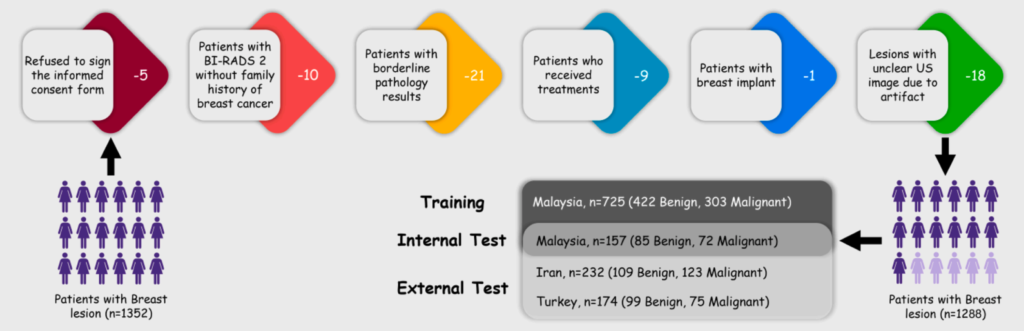
In the present study, three separate data cohorts containing 1288 breast lesions from three countries (Malaysia, Iran, and Turkey) were utilized for MLmodel development and external validation. The model was trained on ultrasound images of 725 breast lesions, and validation was done separately on the remaining data. An expert radiologist and a radiology resident classified the lesions based on the BI-RADS lexicon. Thirteen morphometric features were selected from a contour of the lesion and underwent a three-step feature selection process. Five features were chosen to be fed into the model separately and combined with the imaging signs mentioned in the BI-RADS reference guide. A support vector classifier was trained and optimized.
Results
The diagnostic profile of the model with various input data was compared to the expert radiologist and radiology resident. The agreement of each approach with histopathologic specimens was also determined. Based on BI-RADS and morphometric features, the model achieved an area under the receiver operating characteristic (ROC) curve (AUC) of 0.885, which is higher than the expert radiologist and radiology resident performances with AUC of 0.814 and 0.632, respectively in all cohorts. DeLong’s test also showed that the AUC of the ML protocol was significantly different from that of the expert radiologist (ΔAUCs = 0.071, 95%CI: (0.056, 0.086), P = 0.005).
Conclusions
These results support the possible role of morphometric features in enhancing the already well-excepted classification schemes.
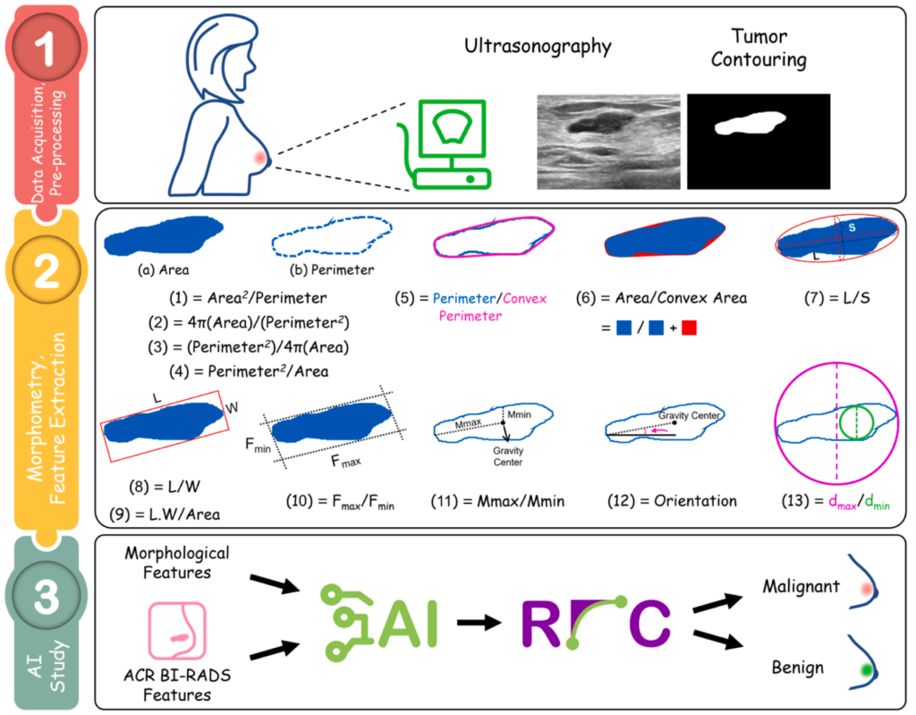
morphometric features of breast lesions; and Step 3, BI-RADS features extraction and ML study.
