Abstract
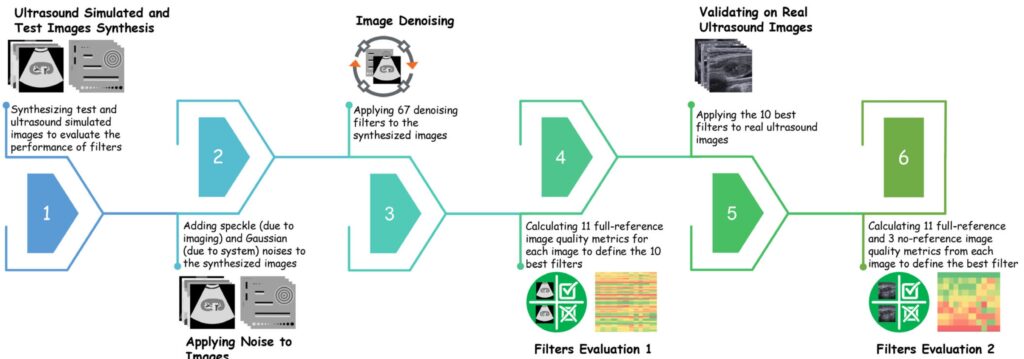
Noise corrupts ultrasound images and degrades spatial and contrast resolutions. Hence, it is challenging to characterize the lesions precisely using ultrasound images. The present study aims to evaluate 67 denoising filters and select the best one for ultrasound image denoising. Seven test images were synthesized to evaluate the performance of filters at three different noise levels. Eleven full-reference quantitative image quality metrics (IQMs) were employed to evaluate the performance of the filters. A new filter evaluation method, Rank Analysis, was introduced and utilized at each noise level. The ten best filters with the smallest mean rank in all noise levels were defined for further analysis on real ultrasound images. The Rank Analysis was also employed for real ultrasound images, and filters were evaluated based on 14 IQMs (11 full-reference and three no-reference). Finally, the best filter was defined using the repeated measures analysis statistical test. According to the Rank Analysis results, the Spatial correlation (SCorr) filter obtained the best results with the mean rank scores±SD of 1 ± 0, which was significantly better than the other nine filters (p < 0.001). The second-best results were achieved by three filters, Bitonic, most homogeneous neighborhood, and Lee diffusion (p < 0.05). We concluded that SCorr is the best filter for ultrasound image denoising. It can be used in the pre-processing step before segmentation and diagnostic procedures. In addition, a new filter evaluation method, Rank Analysis, was introduced in this study, which is easy to use, fast, and provides reliable results. So, it can be used to evaluate newly developed filters in the future studies.