Abstract
Purpose
Our purpose is to assess the inter-rater agreement between digital mammography (DM) and contrast-enhanced spectral mammography (CESM) in evaluating the Breast Imaging Reporting and Data System (BI-RADS) grading.
Approach
This retrospective study included 326 patients recruited between January 2019 and February 2021. The study protocol was pre-registered on the Open Science Framework platform. Two expert radiologists interpreted the CESM and DM findings. Pathological data are used for radiologically suspicious or malignant-appearing lesions, whereas follow-up was considered the gold standard for benign-appearing lesions and breasts without lesions.
Results
For intra-device agreement, both imaging modalities showed “almost perfect” agreement, indicating that different radiologists are expected to report the same BI-RADS score for the same image. Despite showing a similar interpretation, a paired t-test showed significantly higher agreement for CESM compared with DM (p<0.001). Subgrouping based on the side or view did not show a considerable difference for both imaging modalities. For inter-device agreement, “almost perfect” agreement was also achieved. However, for proven malignant lesions, an overall higher BI-RADS score was achieved for CESM, whereas for benign or normal breasts, a lower BI-RADS score was reported, indicating a more precise BI-RADS classification for CESM compared with DM.
Conclusions
Our findings demonstrated strong agreement among readers regarding the identification of DM and CESM findings in breast images from various views. Moreover, it indicates that CESM is equally precise compared with DM and can be used as an alternative in clinical centers.
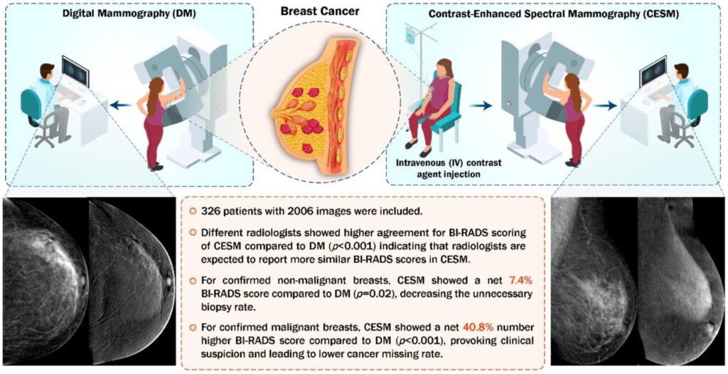
Graphical summary of study findings.
