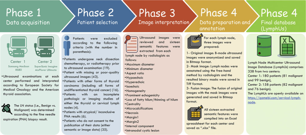
Abstract
Approximately 30–50% of Papillary thyroid carcinoma (PTC) patients develop cervical lymph nodes (LNs) metastasis, significantly increasing the risk of disease recurrence and impacting long-term outcomes. We introduced an open-access multicenter lymph node ultrasound image database (LymphUs) specifically designed to advance research in LN assessment for PTC. Ultrasound imaging was performed on PTC patients at two independent clinical centers using standardized acquisition protocols. Experienced radiologists at each center documented sixteen semantic features for each LN. All LNs were annotated with segmentation masks serving as ground truth, and classification into benign or malignant categories was confirmed by fine needle aspiration biopsy results. The LymphUs comprises ultrasound images with segmentation masks from 338 PTC patients with suspected LN metastasis, divided into two center-specific cohorts: 180 patients (81 malignant, 99 benign) and 158 patients (82 malignant, 76 benign). The complete dataset, including semantic features and expert annotations, is freely accessible for research purposes. The LymphUs bridges a critical gap in medical imaging resources by providing a large-scale, multicenter ultrasound database for cervical LN assessment in PTC, supporting diagnostic algorithms, standardized reporting systems, and artificial intelligence applications to enhance preoperative LN staging and treatment planning.