Abstract
Background:
During the past decade, coronary computed tomographic angiography (CCTA) has become the primary non-invasive imaging technique for the assessment of myocardial bridging (MB).
Objectivs:
The aim of this study was to evaluate the ability of CCTA to predict myocardial ischemia in patients with MB.
Patients and Methods:
A total of 32 MB patients (21 males and 11 females) participated in this study. Eleven MB parameters were measured to assess the ability of CCTA to predict MB patients with ischemia. In order to evaluate ischemia, all the patients underwent single positron emission computed tomography-myocardial perfusion imaging (SPECT-MPI) examination.
Results:
Ischemia was observed in 17 patients (53.1%), while 15 patients (46.9%) did not show signs of ischemia. Out of the 32 patients, superficial MB was observed in 15 patients while deep MB was identified in 12, and borderline was observed in five patients. All MB examined parameters were found to be significantly different between ischemic and non-ischemic patients, except for the location and tunnel artery diameter in diastole. Moreover, a cut-off value of 0.65 mm was able to discriminate ischemia with a sensitivity of 100%, specificity of 93%, and yield area under the receiver operating characteristic (ROC) curve (AUC) of 0.996. Also, by considering the depth cut-off value of 1.75 mm, ischemia can be distinguished with sensitivity and specificity of 100%. MB length had a lower discrimination power, with a cut off value of 22.5 mm yield, 76% sensitivity, 67% specificity, and AUC = 0.810 in the diagnosis of ischemia.
Conclusion:
CCTA was a reliable modality with high accuracy to depict MB, identify high risk MB, and prevent unnecessary SPECT-MPI examination.
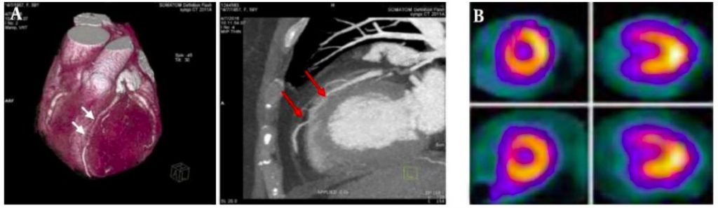
somatom force CT Angiography (A) in 59 years old female with atypical chest pain. The myocardial bridging (MB) is located at mid to distal segment of LAD in length of 20
mm and depth of 2.1 mm (two arrows indicate entrance and exit site of MB). Two selected short and long axis images of the gated SPECT (B) shows reversible perfusion defect
in the mid anteroseptal segments.
