Abstract
Objectives
This study investigated the potential of a clinical decision support approach for the classification of metastatic and tumor-free cervical lymph nodes (LNs) in papillary thyroid carcinoma on the basis of radiologic and textural analysis through ultrasound (US) imaging.
Methods
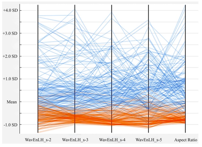
In this research, 170 metastatic and 170 tumor-free LNs were examined by the proposed clinical decision support method. To discover the difference between the groups, US imaging was used for the extraction of radiologic and textural features. The radiologic features in the B-mode scans included the echogenicity, margin, shape, and presence of microcalcification. To extract the textural features, a wavelet transform was applied. A support vector machine classifier was used to classify the LNs.
Results
In the training set data, a combination of radiologic and textural features represented the best performance with sensitivity, specificity, accuracy, and area under the curve (AUC) values of 97.14%, 98.57%, 97.86%, and 0.994, respectively, whereas the classification based on radiologic and textural features alone yielded lower performance, with AUCs of 0.964 and 0.922. On testing the data set, the proposed model could classify the tumor-free and metastatic LNs with an AUC of 0.952, which corresponded to sensitivity, specificity, and accuracy of 93.33%, 96.66%, and 95.00%.
Conclusions
The clinical decision support method based on textural and radiologic features has the potential to characterize LNs via 2-dimensional US. Therefore, it can be used as a supplementary technique in daily clinical practice to improve radiologists’ understanding of conventional US imaging for characterizing LNs.