Abstract
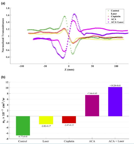
The current chemotherapy method demonstrates the need for improvement in terms of efficacy and safety. Given the beneficiary effect of heat in combination with chemotherapy, the purpose of this study is to develop a multifunctional nanoplatform by co-incorporating gold nanoparticles (AuNPs) as photothermal agent and cisplatin as anticancer drug into alginate hydrogel (named as ACA) to enable concurrent thermo-chemotherapy. The in vitro cytotoxicity experiment showed that the as-developed nanocomplex was able to induce greater cytotoxicity in KB human nasopharyngeal cancer cells compared to free cisplatin at the same concentration. Moreover, the interaction of ACA and laser irradiation acted synergistically and resulted in higher cell death rate compared to separate application of photothermal therapy and chemotherapy. The micrograph of KB cells also revealed that ACA was able to selectively accumulate into the mitochondria, so that laser irradiation of KB cells pre-treated with ACA resulted in intensive morphological damages such as plasma membrane disruption, chromatin condensation, autophagic vacuoles formation and organelle degeneration. Moreover, the sign and magnitude of optical nonlinear refractive index measured by Z-scan technique was shown to be significantly altered in cells exposed to ACA with and without laser irradiation. Consequently, the nanocomplex developed herein could be a promising platform to combine photothermal therapy and chemotherapy effectively, thereby achieving synergistic therapeutic outcome.