Abstract
Background: Cardiac echocardiography and cardiac ECG-gated single-photon emission computed tomography (SPECT) are the most common modalities for left ventricle (LV) volumes and function assessment. The temporal resolution of SPECT images is limited and an ECG provides better temporal resolution. This study investigates the impact of frame numbers on images in terms of qualitative and quantitative assessments.
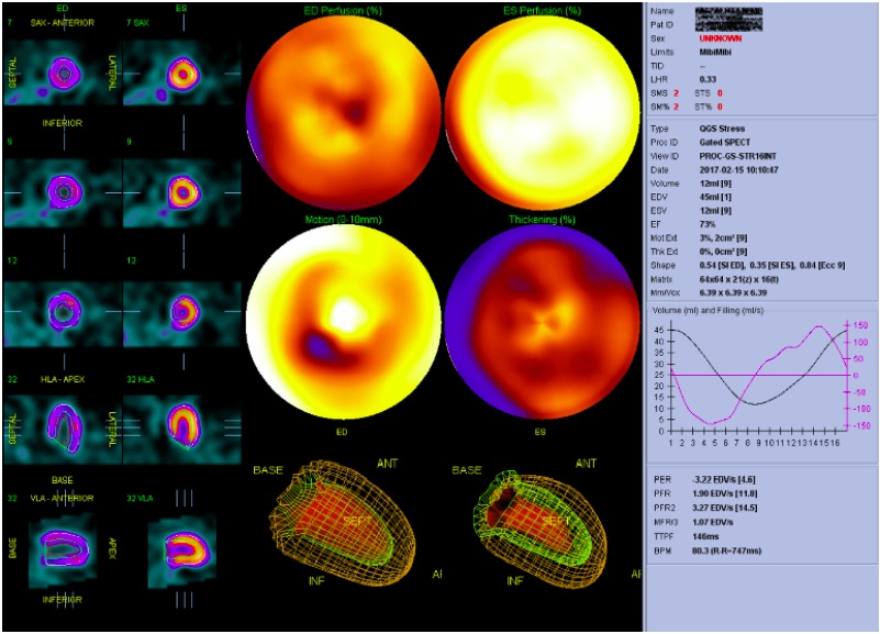
Methods: In this study, 5 patients underwent echocardiography and cardiac ECG-gated SPECT imaging, and 5 standard views of the LV were recorded to determine LV walls boundaries and volumes. Also, 2 original images with 8 frames and 16 frames per cardiac cycle were recorded simultaneously in a single gantry orbit. Using the data extracted from the LV model, 8 extra new frames were created with interpolation between existing frames of the original 8-frame image. Three series of images (8 and 16 original and 16 interpolated) were reconstructed separately. LV volumes and ejection fraction (EF) were calculated using Quantitative Gated SPECT (QGS) software.
Results: Compared to the original 8-frame gating, original 16-frame gated images resulted in larger end-diastole volume (EDV) (mean ± SD: 68.6 ± 27.11 mL vs 66.2±25.41 mL, p<0.001), smaller end-systole volume (ESV) (mean ± SD: 24.6±8.7 mL vs 26±7.3 mL, p<0.001), and higher EF (64% vs 60.2%, p<0.001). The results for the interpolated series were also different from the original images (closer to the original 16-frame series rather than 8-frame).
Conclusion: Changing the frame number from 8 to 16 in cardiac ECG-gated SPECT images caused a significant change in LV volumes and EF. Frame interpolation with sophisticated algorithms can be used to improve the temporal resolution of SPECT images.


