Abstract
Purpose
CT imaging has been a detrimental tool in the diagnosis of COVID-19, but it has not been studied thoroughly in pediatric patients and its role in diagnosing COVID-19.
Methods
27 pediatric patients with COVID-19 pneumonia were included. CT examination and molecular assay tests were performed from all participants. A standard checklist was utilized to extract information, and two radiologists separately reviewed the CT images.
Results
The mean age of patients was 4.7 ± 4.16 (mean ± SD) years. Seventeen patients were female, and ten were male. The most common imaging finding was ground-glass opacities followed by consolidations. Seven patients had a single area of involvement, five patients had multiple areas of involvement, and four patients had diffuse involvement. The sensitivity of CT imaging in diagnosing infections was 66.67%. Also, some uncommon imaging findings were seen, such as a tree-in-bud and lung collapse.
Conclusion
CT imaging shows less involvement in pediatric compared to adult patients, due to pediatric patients having a milder form of the disease. CT imaging also has a lower sensitivity in detecting abnormal lungs compared to adult patients. The most common imaging findings are ground-glass opacities and consolidations, but other non-common imaging findings also exist.
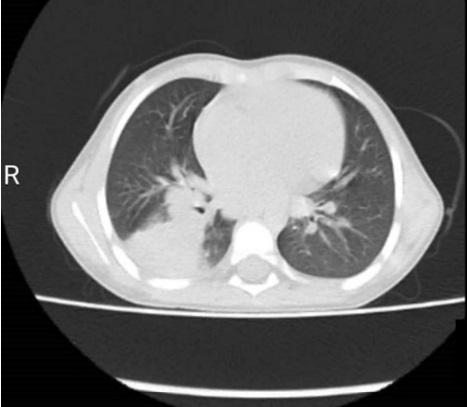
boy shows segmental airspace consolidation of the upper segment of
the right lower lobe
