Abstract
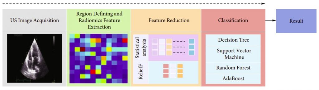
Cardiac pacemakers are used in the treatment of patients with symptomatic bradycardia. The pacemaker paces the heart at the predetermined rate to maintain uninterrupted cardiac activity. Usually, pacemaker lead will be connected to the right atrium (RA) and right ventricle (RV) in dual-chamber pacemaker implantation and RV alone in single-chamber pacemaker implantation. This alters the route of proper conduction across the myocardial cells. The cell-to-cell conduction transmission in pacing delays the activation of selected intraventricular myocardial activation. Pacing-induced cardiomyopathy (PICM) is most commonly defined as a drop in left ventricle ejection fraction (LVEF) in the setting of chronic, high-burden right ventricle (RV) pacing. Currently, very few effective treatments are standard for PICM which rely on the detection of the RV pacing. Such treatments have primarily focused on upgrading to cardiac resynchronization therapy (CRT) when LVEF has dropped. However, the early and accurate detection of these stress factors is challenging. Cardiac desynchrony and interventricular desynchrony can be determined by various echocardiographic techniques, including M-mode, Doppler method, tissue Doppler method, and speckle tracking echocardiography which is subjective measures and shows a significant difference between RV and LV preejection period where the activation of LV is delayed considerably. Computer-aided diagnosis (CAD) is a noninvasive technique that can classify the ultrasound images of the heart in pacemaker-implanted patients and healthy patients with normal left ventricular systolic function and further detect the variations in pacemaker functions in its early stage using heart ultrasound images. Developing such a system requires a vast and diverse database to reach optimum performance. This paper proposes a novel CAD tool for the accurate detection of pacemaker variations using machine learning models of decision tree, SVM, random forest, and AdaBoost. The models have been used to extract radiomics features in terms of textures and then screened by their Relief-F scores for selection and ranking to be classified into nine groups consisting of up to 250 radiomics features. Ten best features were fed to the machine learning models. The R-wave dataset achieved a maximum test performance accuracy of 97.73% with four features in the random forest model. The T-wave dataset achieved a maximum test performance accuracy of 96.59% with three features in the SVM model. Our experimental results demonstrate the system’s robustness, which can be developed as an early and accurate detection system for pacing-induced cardiomyopathy.