Abstract
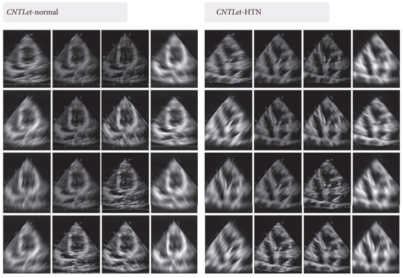
Hypertension (HTN) is a major risk factor for cardiovascular diseases. At least 45% of deaths due to heart disease and 51% of deaths due to stroke are the result of hypertension. According to research on the prevalence and absolute burden of HTN in India, HTN positively correlated with age and was present in 20.6% of men and 20.9% of women. It was estimated that this trend will increase to 22.9% and 23.6% for men and women, respectively, by 2025. Controlling blood pressure is therefore important to lower both morbidity and mortality. Computer-aided diagnosis (CAD) is a noninvasive technique which can determine subtle myocardial structural changes at an early stage. In this work, we show how a multi-resolution analysis-based CAD system can be utilized for the detection of early HTN-induced left ventricular heart muscle changes with the help of ultrasound imaging. Firstly, features were extracted from the ultrasound imagery, and then the feature dimensions were reduced using a locality sensitive discriminant analysis (LSDA). The decision tree classifier with contourlet and shearlet transform features was later employed for improved performance and maximized accuracy using only two features. The developed model is applicable for the evaluation of cardiac structural alteration in HTN and can be used as a standalone tool in hospitals and polyclinics.