Abstract
Purpose
Ultrasonography is the most common imaging modality used to diagnose carpal tunnel syndrome (CTS). Recently artificial intelligence algorithms have been used to diagnose musculoskeletal diseases accurately without human errors using medical images. In this work, a computer-aided diagnosis (CAD) system is developed using radiomics features extracted from median nerves (MN) to diagnose CTS accurately.
Method
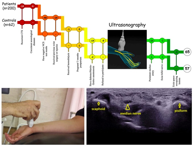
This study is performed on 228 wrists from 65 patients and 57 controls, with an equal number of control and CTS wrists. Nerve conduction study (NCS) is considered as the gold standard in this study. Two radiologists used two guides to evaluate and categorize the pattern and echogenicity of MNs. Radiomics features are extracted from B-mode ultrasound images (Ultrasomics), and the robust features are fed into support vector machine classifier for automated classification. The diagnostic performances of two radiologists and the CAD system are evaluated using ROC analysis.
Results
The agreement of two radiologists was excellent for both guide 1 and 2. The honey-comb pattern clearly appeared in control wrists (based on guide 1). In addition, CTS wrists indicated significantly lower number of fascicles in MNs (based on guide 2). The area under ROC curve (AUC) of the radiologist 1 and 2 are 0.658 and 0.667 based on guide 1 and 0.736 and 0.721 based on guide 2, respectively. The CAD system indicated higher performance than two radiologists with AUC of 0.926.
Conclusion
The proposed CAD system shows the benefit of using ultrasomics features and can assist radiologists to diagnose CTS accurately.