An open-access breast lesion ultrasound image database‏: Applicable in artificial intelligence studies

The current database contains ultrasound images and radiologist-defined masks of two sets of histologically proven benign and malignant lesions...

Abstract

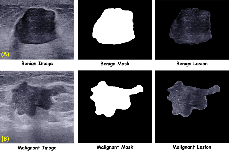
Breast cancer is one of the largest single contributors to the burden of disease worldwide. Early detection of breast cancer has been shown to be associated with better overall clinical outcomes. Ultrasonography is a vital imaging modality in managing breast lesions. In addition, the development of computer-aided diagnosis (CAD) systems has further enhanced the importance of this imaging modality. Proper development of robust and reproducible CAD systems depends on the inclusion of different data from different populations and centers to considerate all variations in breast cancer pathology and minimize confounding factors. The current database contains ultrasound images and radiologist-defined masks of two sets of histologically proven benign and malignant lesions. Using this and similar pieces of data can aid in the development of robust CAD systems.

Sample ultrasound image of benign (A) and malignant (B) breast lesion, and their corresponding mask and resulted fusion images.
Picture of Ardakani AA

Ardakani AA

He received his Ph.D. in Medical Physics in 2018 from the Iran University of Medical Sciences (IUMS), specializing in medical imaging and using artificial intelligence in radiological diagnosis. His research interests focus on the physics of medical imaging systems, quantitative analysis of medical images, and applying artificial intelligence in diagnostic radiology procedures. He is an assistant professor of Medical Physics at Shahid Beheshti University of Medical Sciences.

Leave a Reply

Your email address will not be published. Required fields are marked *