Abstract
INTRODUCTION: Visual inspection by magnetic resonance (MR) images cannot detect microscopic tissue changes occurring in MS in normal appearing white matter (NAWM) and may be perceived by the human eye as having the same texture as normal white matter (NWM). The aim of the study was to evaluate computer aided diagnosis (CAD) system using texture analysis (TA) in MR images to improve accuracy in identification of subtle differences in brain tissue structure.
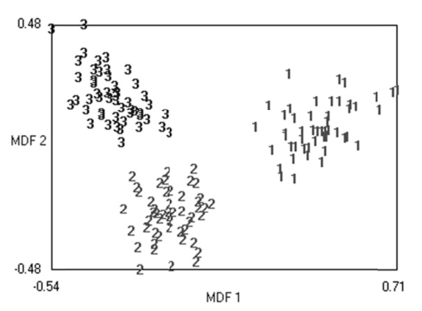
MATERIAL & METHODS: The MR image database comprised 50 MS patients and 50 healthy subjects. Up to 270 statistical texture features extract as descriptors for each region of interest. The feature reduction methods used were the Fisher method, the lowest probability of classification error and average correlation coefficients (POE+ACC) method and the fusion Fisher plus the POE+ACC (FFPA) to select the best, most effective features to differentiate between MS lesions, NWM and NAWM. The features parameters were used for texture analysis with principle component analysis (PCA) and linear discriminant analysis (LDA). Then first nearest-neighbour (1-NN) classifier was used for features resulting from PCA and LDA. Receiver operating characteristic (ROC) curve analysis was used to examine the performance of TA methods.
RESULTS: The highest performance for discrimination between MS lesions, NAWM and NWM was recorded for FFPA feature parameters using LDA; this method showed 100% sensitivity, specificity and accuracy and an area of = 1 under the ROC curve.
CONCLUSION: TA is a reliable method with the potential for effective use in MR imaging for the diagnosis and prediction of MS.