Abstract
Introduction. Ultrasonography is the preferable imaging technique for monitoring and assessing complications in kidney allograft transplants. Computer-aided diagnostic system based on texture analysis in ultrasonographic imaging is recommended to identify changes in kidney function after allograft transplantation.
Materials and Methods. A total of 61 biopsy-proven kidney allograft recipients (11 rejected and 50 unrejected) were assessed by a computer-aided diagnostic system. Up to 270 statistical texture features were extracted as descriptors for each region of interest in each recipient. Correlations of texture features with serum creatinine level and differences between rejected and unrejected allografts were analyzed. An area under the receiver operating characteristic curve was calculated for each significant texture feature. Linear discriminant analysis was employed to analyze significant features and increase discriminative power. Recipients were classified by the first nearest neighbor classifier.
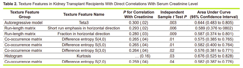
Results. Fourteen texture features had a significant correlation with serum creatinine level and 16 were significantly different between the rejected and unrejected allografts, for which an area under the curve values were in the range of 0.575 for difference entropy S(4,0) to 0.676 for kurtosis. Using all 16 features, linear discriminant analysis indicated higher performance for classification of the two groups with an area under the curve of 0.975, which corresponded to a sensitivity of 90.9%, a specificity of 100%, a positive predictive value of 100%, and a negative predictive value of 98.0%.
Conclusions. Texture analysis was a reliable method, with the potential for characterization, and can help physicians to diagnose kidney failure after transplantation on ultrasonographic imaging.