The LymphUs comprises ultrasound images with segmentation masks from 338 PTC patients with suspected LN metastasis, divided into two center-specific cohorts: 180 patients (81 malignant, 99 benign) and 158 patients (82 malignant, 76 benign). The complete dataset, including semantic features and expert annotations, is freely accessible for research purposes. The LymphUs bridges a critical gap in medical imaging resources by providing a large-scale, multicenter ultrasound database for cervical LN assessment in PTC, supporting diagnostic algorithms, standardized reporting systems, and artificial intelligence applications to enhance preoperative LN staging and treatment planning.
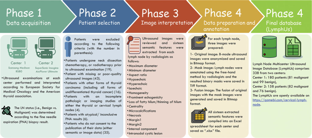
Overall phases involved in this study to generate the LymphUs.
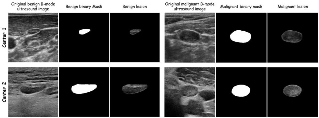
Sample images of benign (First row) and malignant (second row) lymph nodes with the corresponding masks and fused images for both centers.
If you use this database in your research, please cite the following articles:
1- Abbasian Ardakani A, Mohammadi A, Mirza‐Aghazadeh‐Attari M, Faeghi F, Vogl TJ, Acharya UR. Diagnosis of Metastatic Lymph Nodes in Patients With Papillary Thyroid Cancer: A Comparative Multi‐Center Study of Semantic Features and Deep Learning‐Based Models. Journal of Ultrasound in Medicine. 2023 Jun;42(6):1211-21. https://doi.org/10.1002/jum.16131
2- Mohammadi A, Mohebbi A, Mirza-Aghazadeh-Attari M, Mohammadzadeh S, Acharya UR, Tan R-S, et al. LymphUs: A multicenter open-access database of lymph node ultrasound images in patients with papillary thyroid carcinoma for clinical and artificial intelligence research. Data in Brief. 2026:112694. https://doi.org/10.1016/j.dib.2026.112694
