Abstract
Objectives
Ultrasound is widely used in diagnosing carpal tunnel syndrome (CTS). However, the limitations of ultrasound in CTS detection are the lack of objective measures in the detection of nerve abnormality and the operator-dependent nature of ultrasound imaging. Therefore, in this study, we developed and proposed externally validated artificial intelligence (AI) models based on deep-radiomics features.
Methods
We have used 416 median nerves from 2 countries (Iran and Colombia) for the development (112 entrapped and 112 normal nerves from Iran) and validation (26 entrapped and 26 normal nerves from Iran, and 70 entrapped and 70 normal nerves from Columbia) of our models. Ultrasound images were fed to the SqueezNet architecture to extract deep-radiomics features. Then a ReliefF method was used to select the clinically significant features. The selected deep-radiomics features were fed to 9 common machine-learning algorithms to choose the best-performing classifier. The 2 best-performing AI models were then externally validated.
Results
Our developed model achieved an area under the receiver operating characteristic (ROC) curve (AUC) of 0.910 (88.46% sensitivity, 88.46% specificity) and 0.908 (84.62% sensitivity, 88.46% specificity) with support vector machine and stochastic gradient descent (SGD), respectively using the internal validation dataset. Furthermore, both models consistently performed well in the external validation dataset, and achieved an AUC of 0.890 (85.71% sensitivity, 82.86% specificity) and 0.890 (84.29% sensitivity and 82.86% specificity), with SVM and SGD models, respectively.
Conclusion
Our proposed AI models fed with deep-radiomics features performed consistently with internal and external datasets. This justifies that our proposed system can be employed for clinical use in hospitals and polyclinics.
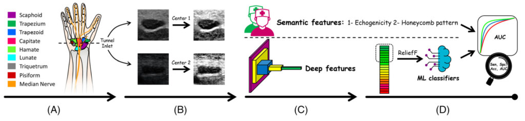
Graphical abstract of the study. Overview of the 4 steps used to differentiate carpal tunnel syndrome from normal wrists. A, Ultrasound imaging at the tunnel inlet. B, Preprocessing, image filtering. C, Feature extraction. D, Feature analysis and classification.
