Abstract
Purpose: Hyperthermia and radiation have the ability to induce structural and morphological changes on both macroscopic and microscopic level. Normal and damage cells have a different texture but may be perceived by human eye, as having the same texture.
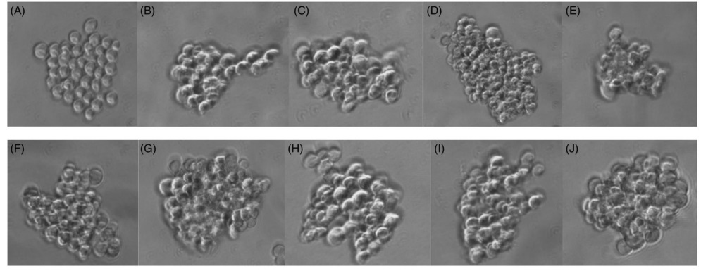
Materials and methods: To explore the potential of texture analysis based on run-length matrix, a total of 32 sphere images for each group and treatment regime were used in this study. Cells were subjected to the treatment with different doses of 6 MeV electron radiation (0 2, 4 and 6 Gy), hyperthermia (at 43° C in 0, 30, 60 and 90 min) and radiation + hyperthermia (at 43 °C in 30 min with 2, 4 and 6 Gy dose), respectively. Twenty run-length matrix (RLM) features were extracted as descriptors for each selected region of interest for texture analysis. Linear discriminant analysis was employed to transform raw data to lower-dimensional spaces and increase discriminative power.
Results: The features were classified by the first nearest neighbor classifier. RLM features represented the best performance with sensitivity, specificity, accuracy, positive predictive value (PPV) and negative predictive value (NPV) of 100% between 0 and 6 Gy radiation, 0 and 6 Gy radiation + hyperthermia, 0 and 90 min and 30 and 90 min hyperthermia groups. The area under receiver operating characteristic curve was 1 for these groups.
Conclusion: RLM features have a high potential to characterize cell changes during different treatment regimes.