Diagnosis of Metastatic Lymph Nodes in Patients with Papillary Thyroid Cancer: A Comparative Multi-Center Study of Semantic Features and Deep Learning-Based Models

In the present study, we report the diagnostic profile of a deep learning model in differentiating malignant and benign lymph nodes in patients with papillary thyroid cancer...

Abstract

Objectives

Deep learning algorithms have shown potential in streamlining difficult clinical decisions. In the present study, we report the diagnostic profile of a deep learning model in differentiating malignant and benign lymph nodes in patients with papillary thyroid cancer.

Methods

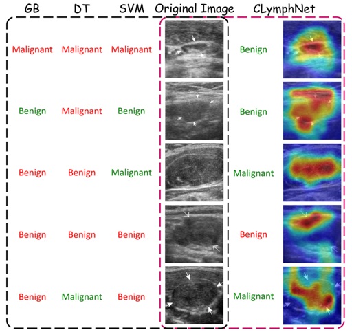
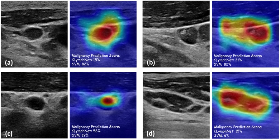
An in-house deep learning-based model called “ClymphNet” was developed and tested using two datasets containing ultrasound images of 195 malignant and 178 benign lymph nodes. An expert radiologist also viewed these ultrasound images and extracted qualitative imaging features used in routine clinical practice. These signs were used to train three different machine learning algorithms. Then the deep learning model was compared with the machine learning models on internal and external validation datasets containing 22 and 82 malignant and 20 and 76 benign lymph nodes, respectively.

Results

Among the three machine learning algorithms, the support vector machine model (SVM) outperformed the best, reaching a sensitivity of 91.35%, specificity of 88.54%, accuracy of 90.00%, and an area under the curve (AUC) of 0.925 in all cohorts. The ClymphNet performed better than the SVM protocol in internal and external validation, achieving a sensitivity of 93.27%, specificity of 92.71%, and an accuracy of 93.00%, and an AUC of 0.948 in all cohorts.

Conclusion

A deep learning model trained with ultrasound images outperformed three conventional machine learning algorithms fed with qualitative imaging features interpreted by radiologists. Our study provides evidence regarding the utility of ClymphNet in the early and accurate differentiation of benign and malignant lymphadenopathy.

Comparison of misclassified lymph nodes by different models for the internal validation dataset. Correctly and incorrectly diagnosed lymph nodes by each model are shown in green and red colors, respectively. The Grad-CAM images of the CLymphNet model are also shown for better visualization (right column).
Original B-mode with the corresponding Grad-CAM images of tumor-free (A, B) and metastatic lymph nodes (C, D). The malignancy prediction scores of the CLymphNet model and the best ML model, SVM, are shown in percentage. (A) The tumor-free node was classified correctly by both models. (B) The tumor-free node was classified correctly and incorrectly by CLymphNet and SVM models, respectively. (C) The metastatic node was classified correctly and incorrectly by CLymphNet and SVM models, respectively. (D) The metastatic node was classified incorrectly by both models.
Picture of Ardakani AA

Ardakani AA

He received his Ph.D. in Medical Physics in 2018 from the Iran University of Medical Sciences (IUMS), specializing in medical imaging and using artificial intelligence in radiological diagnosis. His research interests focus on the physics of medical imaging systems, quantitative analysis of medical images, and applying artificial intelligence in diagnostic radiology procedures. He is an assistant professor of Medical Physics at Shahid Beheshti University of Medical Sciences.

Leave a Reply

Your email address will not be published. Required fields are marked *