Abstract
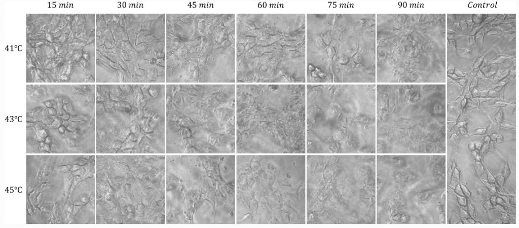
Hyperthermia treatment can induce component changes on cell. This study explored the potential of Z-scan to improve accuracy in the identification of subtle differences in mouse colon cancer cell line CT26 during hyperthermia treatment. Twenty-one samples were subjected individually to treatment of hyperthermia at 41, 43, and 45 °C. Each hyperthermia treatment was done in six different time (15, 30, 45, 60, 75, and 90 min). Two optical setups were used to investigate the linear and nonlinear optical behavior of samples. Prior to the Z-scan technique, all samples were fixed with 1 mL of 5% paraformaldehyde. The linear optical setup indicated that extinction coefficient cannot monitor cell changes at different treatment regimes. But the nonlinear behavior of CT26 in all hyperthermia treatment regimens was different. By increasing the time and/or temperature of hyperthermia treatments, change in the sign of nonlinear refractive index from negative to positive occurred in earlier time intervals. This phenomenon was seen for 41, 43, and 45 °C in 75, 60, and 45 min, respectively. The results showed that the Z-scan technique is a reliable method with the potential to characterize cell changes during hyperthermia treatment regimes. Nonlinear refractive index can be used as a new index for evaluation of cell damage.


