Abstract
Background
Although traditional treatments are able to increase cancer survival rate, undesirable impact on off-target tissues are considered a limitation of these approaches. Nanotechnology-based treatments have been proposed as a possible option to enhance targeting. Further, current methods for evaluating cellular damage, are time consuming, highly dependent on the operator skills, and expensive. The aim of this study was to evaluate the capability of nonlinear optical response of cells to determine cellular damages during conventional and nano-technology based treatments.
Methods
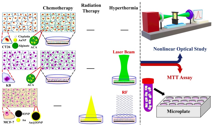
Three different cancer cell lines, CT26, KB, and MCF-7 were used in this study. The alginate hydrogel co-loaded with cisplatin and Au nanoparticle (ACA) nanocomplex and gold-coated iron oxide nanoparticle (Au@IONP) were considered for chemo- and chemo-photothermal therapies, and thermo-radiation therapy, respectively. The sign and value of nonlinear optical absorption coefficient and imaginary part of the third-order nonlinear susceptibility of cells were computed. MTT assay was utilized as a reference method.
Results
The value of nonlinear optical indices increased with increasing cellular damage and cell death. The linear regression analysis indicated high correlation between nonlinear optical indices and MTT results, in all treatments.
Conclusion
The nonlinear optical indices are robust from confounding factors, namely treatment approach (traditional and nano-technology based), treatment modality (chemotherapy, thermotherapy, photothermal therapy, and radiation therapy), and cell types. Nonlinear optical properties of cells can be used as a rapid estimation method for cell damage, at the nanoscale level.