Abstract
Introduction: Assessing local invasion is essential for determining stage of cN0 head and neck squamous cell carcinoma (HNSCC). We aimed to evaluate the performance of fluorine-18 fluorodeoxyglucose positron emission tomography/CT (18F-FDG PET/CT) in HNSCC characterization and compare it with conventional imaging.
Methods: This multicentral study included 278 consecutive newly diagnosed cN0 HNSCC patients recruited from ACRIN 6685 (American College of Radiology Imaging Network) dataset. Four boardcertified nuclear radiologists interpreted preoperative PET/CT, MRI, and CT examinations of patients. Imaging results were compared to pathological reference tests through area under curve (AUC), sensitivity, specificity, and receiver operating characteristic (ROC) curve using Stata 18 and Medcalc 22.017.
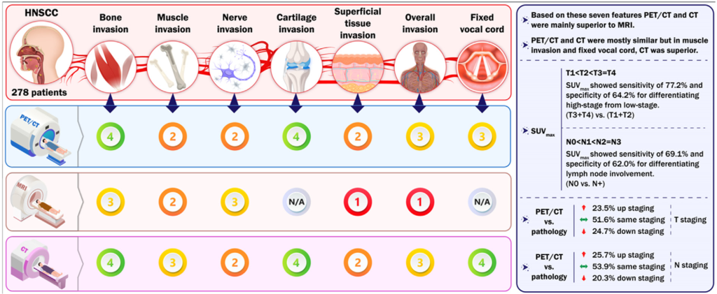
Results: PET/CT demonstrated 23.5 %, 24.7 %, and 51.6 % upstaging, downstaging, and same staging in T staging of patients in comparison to histopathological evaluation, respectively. When evaluating N status, PET/CT showed 25.7 % upstaging, 20.3 % downstaging, and 53.9 % same staging. An optimal SUVmax cutoff value of 10.9 was determined to predict early-stage (T1, T2) and advanced-stage (T3, T4) HNSCC tumors with an AUC of 0.709 (95 % CI ¼ 0.648e0.766). This cut-off value also predicted N0 and Nþ patients with an AUC of 0.670 (95 % CI ¼ 0.606e0.729). Sensitivity and specificity of PET/CT, MRI, and CT for bone invasion, muscle invasion, nerve invasion, cartilage invasion, superficial tissue invasion, overall invasion, and fixed vocal cord were calculated.
Conclusion: Our findings support the valuable accuracy of 18F-FDG PET/CT in staging HNSCC patients. Also, 18F-FDG PET/CT outperformed conventional imaging in characterization of HNSCC tumors. Implications for practice: By offering an in-depth investigation in imaging of HNSCC tumors, this study contributes to evidence-based clinical decision-making.

Figure 2. A visual graph containing critical findings of the study.

