Abstract
Objectives
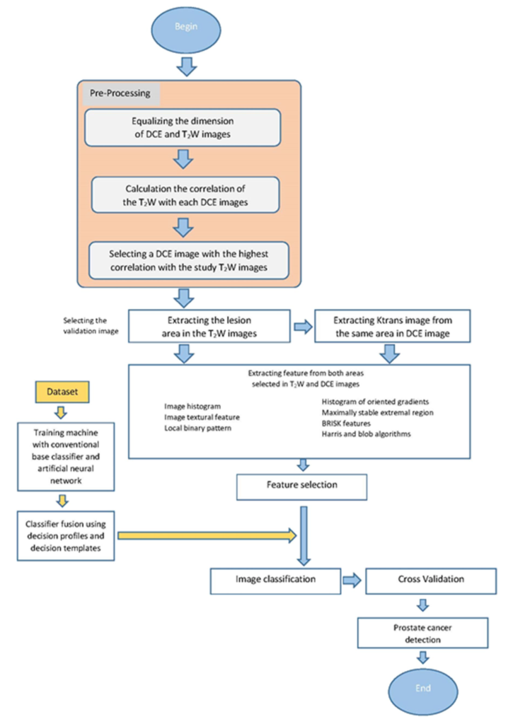
Multiparametric MRI (mp-MRI) has been significantly used for detection, localization and staging of Prostate cancer (PCa). However, all the assessment suffers from poor reproducibility among the readers. The aim of this study was to evaluate radiomics models to diagnose PCa using high-resolution T2-weighted (T2-W) and dynamic contrast-enhanced (DCE) MRI.
Materials and methods
Thirty two patients who had high prostate specific antigen level were recruited. The prostate biopsies considered as the reference to differentiate between 66 benign and 36 malignant prostate lesions. 181 features were extracted from each modality. K-nearest neighbors, artificial neural network, decision tree, and linear discriminant analysis were used for machine-learning study. The leave-one-out cross-validation method was used to prevent overfitting and build robust models.
Results
Radiomics analysis showed that T2-W images were more effective in PCa detection compare to DCE images. Local binary pattern features and speeded up robust features had the highest ability for prediction in T2-W and DCE images, respectively. The classifier fusion using decision template method showed the highest performance with accuracy, specificity, and sensitivity of 100%.
Discussion
The findings of this framework provide researchers on PCa with a promising method for reliable detection of prostate lesions in MR images by fused model.