Abstract
Purpose
Early detection and monitoring of kidney function during the post-transplantation period is one of the most important issues for improving the accuracy of an initial diagnosis. The aim of this study was to evaluate texture analysis (TA) in scintigraphic imaging to detect changes in kidney status after transplantation.
Material and methods
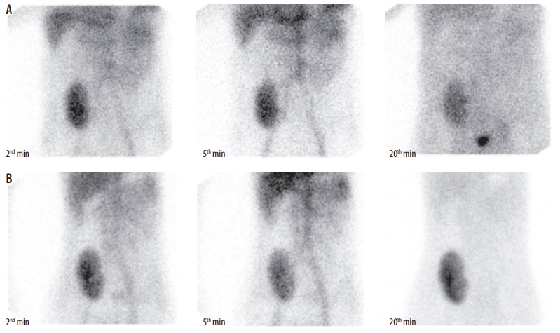
Scintigraphic images were used for TA from a total of 94 kidney allografts (39 rejected and 55 non-rejected). Images corresponding to the frames at the 2nd, 5th, and 20th minute of the study were used to determine the optimum time point for analysis of differences in texture features between the rejected and non-rejected allografts.
Results
Linear discriminant analysis indicated the best performance at the fifth minute frame for classification of the rejected and non-rejected allografts with receiver operating characteristic curve (Az) of 0.982, corresponding to 91.89% sensitivity, 96.49% specificity, and 94.68% accuracy. Also, TA can differentiate acute tubular necrosis from acute rejection with Az of 0.953 corresponding to 88% sensitivity, 92.31% specificity, and 90.62% accuracy at the 5th minute frame. The best correlation between texture feature and kidney function was achieved at the 20th minute frame (r = –0.396) for glomerular filtration rate.
Conclusions
TA has good potential for the characterisation of kidney failure after transplantation and can improve clinical diagnosis.