Abstract
Objectives
The aim of this study was to evaluate if the analysis of sonographic parameters could predict if a thyroid nodule was hot or cold.
Methods
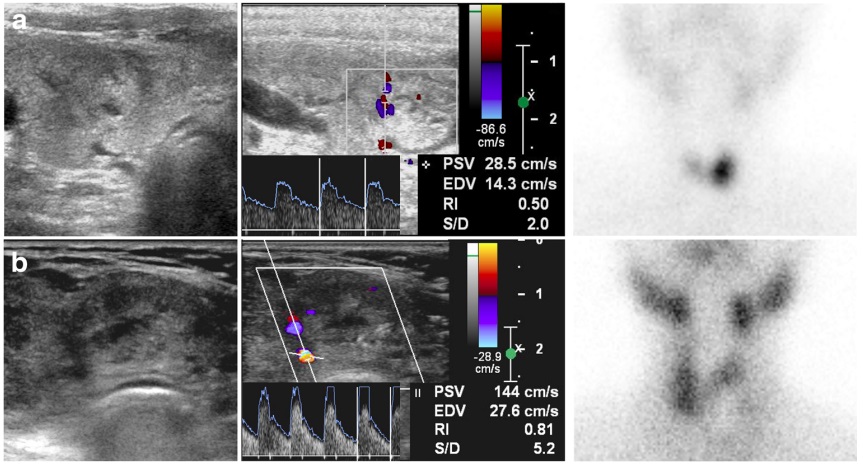
Overall, 102 thyroid nodules, including 51 hyperfunctioning (hot) and 51 hypofunctioning (cold) nodules, were evaluated in this study. Twelve sonographic features (i.e., seven B-mode and five Doppler features) were extracted for each nodule type. The isthmus thickness, nodule volume, echogenicity, margin, internal component, microcalcification, and halo sign features were obtained in the B-mode, while the vascularity pattern, resistive index (RI), peak systolic velocity, end diastolic velocity, and peak systolic/end diastolic velocity ratio (SDR) were determined, based on Doppler ultrasounds. All significant features were incorporated in the computer-aided diagnosis (CAD) system to classify hot and cold nodules.
Results
Among all sonographic features, only isthmus thickness, nodule volume, echogenicity, RI, and SDR were significantly different between hot and cold nodules. Based on these features in the training dataset, the CAD system could classify hot and cold nodules with an area under the curve (AUC) of 0.898. Also, in the test dataset, hot and cold nodules were classified with an AUC of 0.833.
Conclusions
2D sonographic features could differentiate hot and cold thyroid nodules. The CAD system showed a great potential to achieve it automatically.