Abstract
Background
Investigation of cortical bone using magnetic resonance imaging is a developing field, which uses short/ultrashort echo time (TE) pulse sequences to quantify bone water content and to obtain indirect information about bone microstructure.
Purpose
To improve the accuracy of the previously proposed technique of free water T1 quantification and to seek the relationship between cortical bone free water T1 and its mechanical competence.
Study Type
Prospective.
Subjects
Twenty samples of bovine tibia bone.
Field Strength/Sequences
3.0 T; ultra-fast two-dimensional gradient echo, Radio frequency-spoiled three-dimensional gradient echo.
Assessment
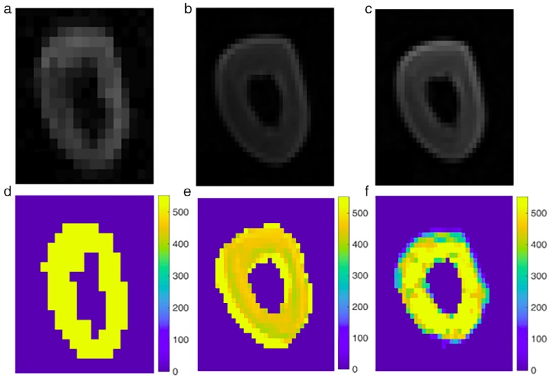
Cortical bone free water T1 was quantified via three different methods: inversion recovery (IR), variable flip angle (VFA), and variable repetition time (VTR). Signal-to-noise ratio was measured by dividing the signal of each segmented sample to background noise. Segmentation was done manually. The effect of noise on T1 quantification was evaluated. Then, the samples were subjected to mechanical compression test to measure the toughness, yield stress, ultimate stress, and Young modulus.
Statistical Tests
All the statistical analysis (Shapiro–Wilk, way analysis of variance, paired t test, Pearson correlation, and Bland–Altman plot) were done using SPSS.
Results
Significant difference was found between T1 quantification groups (P < 0.05). Average T1 of each quantification method differed significantly after adding noise (P < 0.05). VFA-T1 values significantly correlated with toughness (r = −0.68, P < 0.05), ultimate stress (r = −0.71, P < 0.05), and yield stress (r = −0.62, P < 0.05). No significant correlation was found between VTR-T1 values and toughness (P = 0.07), ultimate stress (P = 0.47), yield stress (P = 0.30), and Young modulus (P = 0.39).
Data Conclusion
Pore water T1 value is associated with bone mechanical competence, and VFA method employing short-TE pulse sequence seems a superior technique to VTR method for this quantification.